Fetal Echocardiography in New Delhi – Advanced Assessment of Your Baby’s Heart Before Birth
A Fetal Echocardiography (Fetal Echo) is a specialized ultrasound examination that evaluates the structure and function of a baby’s heart during pregnancy. It helps detect congenital heart abnormalities before birth and allows doctors to closely monitor the baby’s cardiac health. Early diagnosis of heart conditions enables timely medical planning, appropriate treatment, and better outcomes for both the mother and baby.
If you are looking for a trusted Fetal Echocardiography in New Delhi, Dr. Anshu Chamoli provides comprehensive pregnancy care with advanced fetal monitoring as part of personalized antenatal services. With over 10 years of experience in Obstetrics, Gynaecology, and Reproductive Medicine, she ensures timely evaluation and appropriate guidance to support a healthy pregnancy and safe delivery.
What is Fetal Echocardiography?
Fetal Echocardiography, commonly known as a Fetal Echo, is a detailed ultrasound scan specifically designed to examine the baby’s heart before birth. Unlike a routine pregnancy ultrasound, which evaluates overall fetal growth and development, a fetal echo focuses exclusively on the heart’s structure, chambers, valves, blood vessels, rhythm, and blood flow.
This specialized scan helps identify congenital heart defects and other cardiac conditions that may require monitoring during pregnancy or treatment after birth. It is usually performed during the second trimester, although the timing may vary depending on the pregnancy and medical indications.
Why is Fetal Echocardiography Important?
A Fetal Echo provides detailed information about your baby’s heart health and allows early detection of cardiac abnormalities that may not be visible on a routine ultrasound.
Key benefits include:
Detailed assessment of the baby’s heart structure
Evaluation of heart function and rhythm
Early detection of congenital heart defects
Assessment of blood flow through the heart
Monitoring high-risk pregnancies
Better planning for delivery and newborn care
Timely referral for specialized treatment if required
Reassurance when the baby’s heart is developing normally
Early diagnosis helps doctors prepare the most appropriate care plan for the baby before and after birth.
What Happens During Fetal Echocardiography?
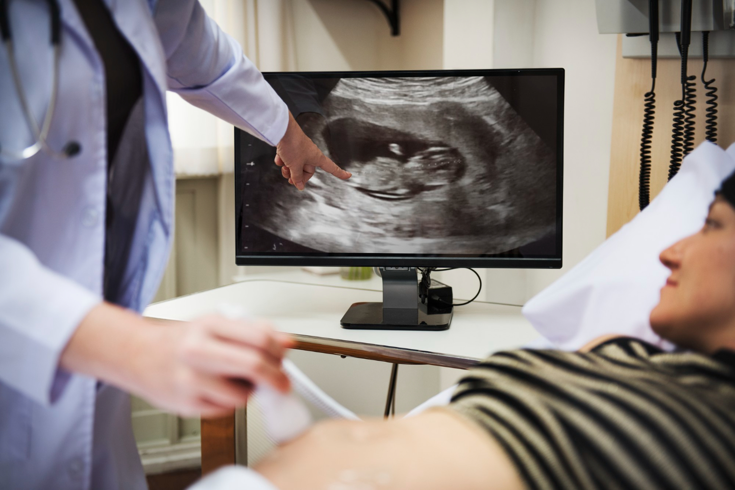
A Fetal Echo is a safe, painless, and non-invasive ultrasound examination. During the procedure, a special gel is applied to the mother’s abdomen, and an ultrasound probe is gently moved over the skin to obtain detailed images of the baby’s heart.
During the scan, the doctor may evaluate:
Heart chambers
Heart valves
Major blood vessels
Heart rhythm
Blood flow through the heart
Heart rate
Overall cardiac structure
Baby’s general well-being
The procedure usually takes between 30 and 60 minutes, depending on the baby’s position and the level of detail required for assessment.
Benefits of Fetal Echocardiography
A Fetal Echo offers several important benefits in monitoring your baby’s heart health during pregnancy.
Early Detection of Heart Conditions
The scan helps identify congenital heart defects before birth, allowing timely medical planning.
Comprehensive Cardiac Assessment
Detailed evaluation of the baby’s heart structure and function provides valuable diagnostic information.
Improved Pregnancy Management
Early diagnosis allows doctors to closely monitor the pregnancy and prepare for specialized care if needed.
Better Delivery Planning
Knowing about a heart condition before birth helps ensure that delivery takes place in an appropriate medical setting with the required support.
Parental Reassurance
When the baby’s heart is developing normally, the scan provides confidence and peace of mind for expecting parents.
Safe and Non-Invasive Procedure
Fetal Echocardiography uses ultrasound technology without radiation, making it safe for both mother and baby.
Who Should Have a Fetal Echocardiography?
A Fetal Echo may be recommended when there is an increased risk of congenital heart disease or when additional evaluation of the baby’s heart is needed.
It is particularly recommended for women who:
Have a family history of congenital heart disease
Have diabetes before pregnancy
Have certain autoimmune disorders
Have conceived through IVF or fertility treatment
Are carrying twins or multiple babies
Have abnormal findings on a routine pregnancy ultrasound
Have certain infections or medical conditions during pregnancy
Are carrying a baby suspected to have a heart abnormality
Your doctor will recommend the scan if it is appropriate based on your pregnancy and medical history.
Why Choose Dr. Anshu Chamoli for Fetal Echocardiography in New Delhi?
Dr. Anshu Chamoli is a highly experienced Consultant Gynaecologist and IVF Specialist committed to providing comprehensive pregnancy care through advanced fetal assessment and personalized treatment. With advanced qualifications in Obstetrics & Gynaecology, Fellowship in Minimal Invasive Gynaecological Surgery (MIGS), and Fellowship in Reproductive Medicine, she ensures that every pregnancy receives careful monitoring using evidence-based clinical practices.
Her patient-centered approach focuses on early diagnosis, detailed pregnancy evaluation, compassionate communication, and individualized care. She works closely with expecting parents to ensure every stage of pregnancy is monitored with accuracy, helping achieve the best possible outcomes for both mother and baby.
If you are searching for a trusted Fetal Echocardiography in New Delhi, consult Dr. Anshu Chamoli for advanced fetal heart assessment, expert pregnancy monitoring, and personalized antenatal care that prioritizes your baby’s heart health and overall well-being.
Frequently Asked Questions (FAQs)
1. What is the purpose of a Fetal Echocardiography?
A Fetal Echo evaluates the structure and function of the baby’s heart and helps detect congenital heart defects before birth.
2. Is Fetal Echocardiography safe during pregnancy?
Yes. It is a safe, non-invasive ultrasound procedure that uses sound waves and does not expose the mother or baby to radiation.
3. When is a Fetal Echo usually performed?
It is commonly performed during the second trimester, although your doctor may recommend it earlier or later depending on your pregnancy.
4. Is a Fetal Echo different from a routine pregnancy ultrasound?
Yes. A routine ultrasound assesses overall fetal growth and anatomy, while a Fetal Echo focuses specifically on the baby’s heart structure, function, and blood flow.
5. Will every pregnant woman need a Fetal Echocardiography?
No. A Fetal Echo is generally recommended when there are specific medical indications, risk factors, or abnormal findings that require a detailed evaluation of the baby’s heart.